Galerie
Slide shows
| Differenziertes Krafttraining | |
| Blum: Kinesiologische Analyse | |
| Unterkiefer | |
| Ren mobilis (Die Wanderniere) |
Schriftliches

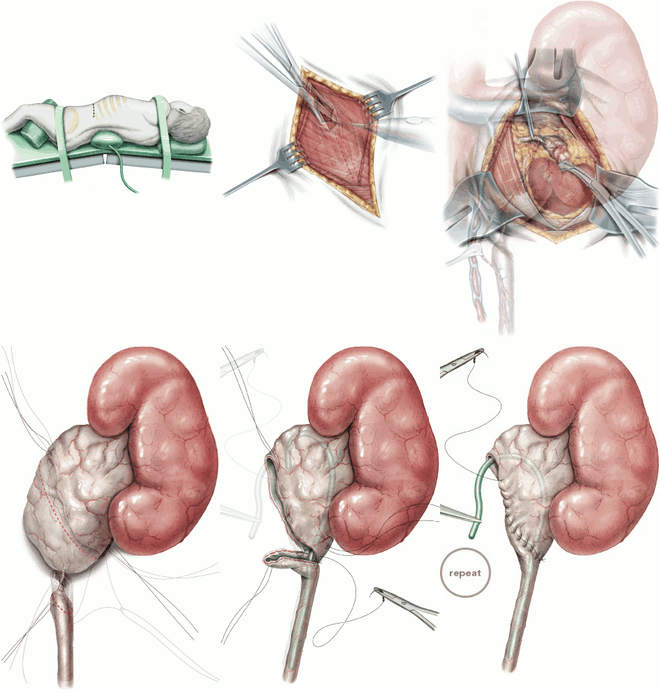
| Title | Pyeloplasty (Anderson-Hynes) |
| Technique | Pencil / Watercolour / Computer |
| Author | Hensle / Shabsigh, Childrens Hospital of New York, NYC |
| Publication | British Journal of Urology International (BJUI) |
| Publisher | Blackwell-Wiley Publishing 2004 |
| close window | |
| Figure 1 | Patient positioning (infant) and skin incision |
| Figure 2 | Incision of the internal and external oblique muscles |
| Figure 3 | Oblique muscles are retracted back, blunt incision of the transversus abdominus muscle |
| Figure 4 | The peritoneum is bluntly displaced medially and lumbodorsal fascia incised |
| Figure 5 | Great care must be taken to minimize trauma and preserve bloody supply in the region of the upper ureter |
| Figure 6 | Fine stay sutures are placed, the incisions are marked |
| Figure 7 | 2-3 cm spatulation of the ureter on its inferior border. |
| Figure 8 | A stent protects the back wall of the ureter from the sutures used for anastomoses |
| Figure 9 | Before the anastomoses is completed the temporary stent is removed |
| close window | |