Galerie
Slide shows
| Differenziertes Krafttraining | |
| Blum: Kinesiologische Analyse | |
| Unterkiefer | |
| Ren mobilis (Die Wanderniere) |
Schriftliches

| Title | Laparoscopic right donor nephrectomy |
| Technique | Pencil / Watercolour / Computer |
| Author | Kapoor/Lambe/Whelan, Mc Master Institute at St. Joseph Healthcare, Ontario, Canada |
| Publication | British Journal of Urology International (BJUI) |
| Publisher | Blackwell-Wiley Publishing 2009 |
| close window | |
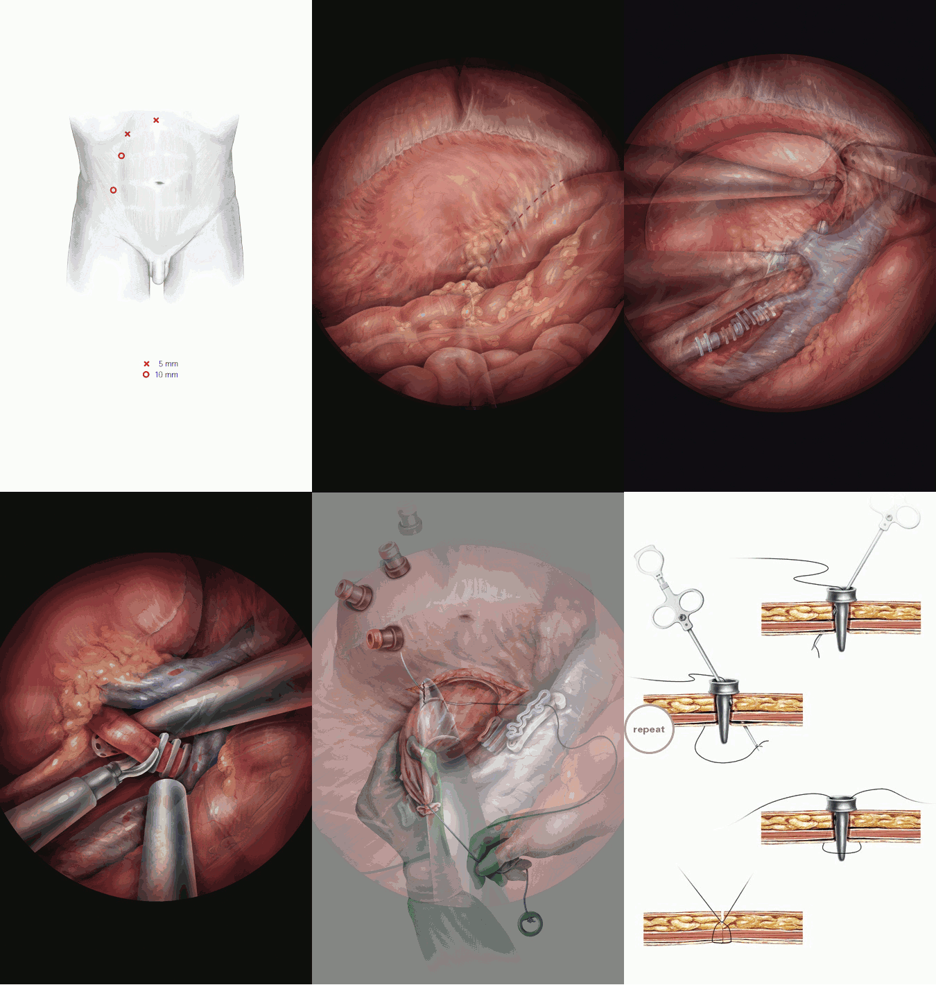
| Figure 1 | Port placement |
| Figure 2 | Right colon mobilization |
| Figure 3 | Right colon mobilization |
| Figure 4 | Gonadal vein clipped and divided |
| Figure 5 | Gerota's fascia is incised and the adrenal gland is dissected off the upper pole of the kidney |
| Figure 6 | Identification and mobilization of the right renal vein |
| Figure 7 | The renal artery is exposed and dissected for an adequate length behind the IVC |
| Figure 8 | The renal artery is occluded with three metal clips and cut |
| Figure 9 | Renal vein clipped (Hem-o-lok) and cut, insertion of the specimen bag |
| Figure 10 | Over-suturing the renal vein stump |
| Figure 11 | Before the kidney was retrieved through a Pfannenstiel incision |
| Figure 12 | Carter Thomason device for 10 mm port closure |
| close window | |