Galerie
Slide shows
| Differenziertes Krafttraining | |
| Blum: Kinesiologische Analyse | |
| Unterkiefer | |
| Ren mobilis (Die Wanderniere) |
Schriftliches

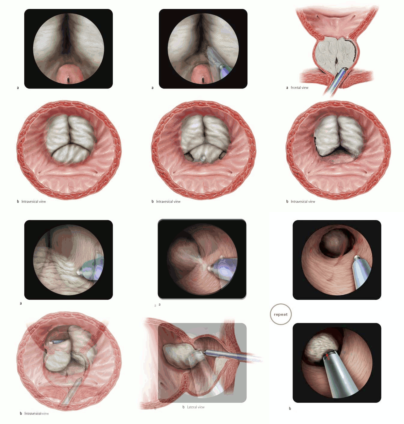
| Title | Holmium Laser Enucleation of the prostate (HoLEP) |
| Technique | Pencil / Watercolour / Computer |
| Author | Prof. Dr. Peter Gilling, Tauranga Hospital, New Zealand |
| Publication | British Journal of Urology International (BJUI) 2008 |
| Publisher | Blackwell-Wiley Publishing 2008 |
| close window | |
| Figure 1 | Preliminary cystoscopy is used to ascertain the size and configuration of the prostate, and to assess the patient for other pathology such as bladder calculi. |
| Figure 2 | Bilateral bladder neck incisions are then made, extending from the ureteric orifices to the verumontanum. |
| Figure 3 | Once the incisions are complete, the median lobe is enucleated starting at the verumontanum. |
| Figure 4 | The lateral lobes are each enucleated in several distinct steps. First, the distal bladder neck incision is extended out inferolaterally ... |
| Figure 5 | ... and then continued upwards at the level of the verumontanum to find the surgical plane and to begin to define the apex. |
| Figure 6 | The lower margin of the lobe is released commencing at the apex, and the apical incision is continued up to the 2 o'clock position. |
| Figure 7 | The upper and lower incisions are connected at the apex and the lateral lobe is enucleated in the capsular plane. |
| Figure 8 | Once the lobes has been placed in the bladder the prostatic fossa is further inspected and any remaining bleeding dealt with. For morcellation (Figure 8b), the inner sheath is replaced by the long nephroscope and adapter. |
| close window | |