Galerie
Slide shows
| Differenziertes Krafttraining | |
| Blum: Kinesiologische Analyse | |
| Unterkiefer | |
| Ren mobilis (Die Wanderniere) |
Schriftliches

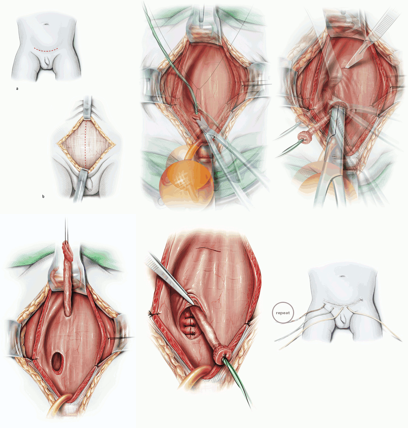
| Title | Politano-Leadbetter ureteric reimplantation |
| Technique | Pencil / Watercolour / Computer |
| Author | Steffens / Treyer / Stark / Haben, St. Antonius Hospital, Eschweiler |
| Publication | British Journal of Urology International (BJUI) |
| Publisher | Blackwell-Wiley Publishing 2006 |
| close window | |
| Figure 1 | Pfannenstiel incision and longitudinal incision of rectus fascia |
| Figure 2 | Longitudinal bladder dome incision |
| Figure 3 | Bilateral fixation of the bladder wall at rectus fascia. Insertion and fixation of an ureteric stent. |
| Figure 4 | Circumferential incision of the ureteric orifice |
| Figure 5 | Distal ureterolysis; the preparation is made close to the ureter in the layer of the meso-ureter. |
| Figure 6 | Transvesical mobilization of the adherent peritoneum from the distal ureter using a Langenbeck retractor for better visualization |
| Figure 7 | Creation of a neo-hiatus |
| Figure 8 | After pereparation of a wide neo-hiatus, grasping of a free suture as a guide rail for the ureter |
| Figure 9 | Retraction of one suture end into the bladder and fixation with the stay suture of the ureteric orifice after stent removal |
| Figure 10 | Passage of the ureter through the neo-hiatus |
| Figure 11 | Transvesical transposition of the distal ureter. Submucosal tunnel preparation from the old to the new hiatus. |
| Figure 12 | Closure of the bladder wall defect using three sutures |
| Figure 13 | Submucosal transposition of the distal ureter |
| Figure 14 | Fixation of the orifice at initial position after resection of a dysplastic ureteric segment. Closure of the bladder mucosa over the reimplanted ureter. |
| Figure 15 | Wound closure. Drainage, ureteric stent and two cystostomies in place. |
| close window | |